Nanomagnetism & Oxides Laboratory
Biomagnetism
People: Myriam Pannetier-Lecoeur, Claude Fermon, Aurélie Solignac, Jacob Torrejon-Diaz (postdoc), Chloé Chopin (PhD)
Alumni: Vincent Trauchessec (PhD), Laure Caruso (PhD), Josué Trejo-Rosillo (Postdoc), Paolo Campilo (Postdoc), Hedwige Polovy (PhD)
Collaborations (current): ESI Frankfurt University of Stuttgart
Fundings (current): ANR, DFG, DRF impulsion
Alumni: Vincent Trauchessec (PhD), Laure Caruso (PhD), Josué Trejo-Rosillo (Postdoc), Paolo Campilo (Postdoc), Hedwige Polovy (PhD)
Collaborations (current): ESI Frankfurt University of Stuttgart
Fundings (current): ANR, DFG, DRF impulsion
In vivo local recording of the magnetic signature of neurons
Neuronal activity generates ionic flows and thereby both magnetic fields and electric potential differences,
i.e., voltages. Voltage measurements are widely used but suffer from isolating and smearing properties of tissue between source and sensor. They are blind to ionic flow direction, and reflect the difference between two electrodes, complicating interpretation. Magnetic field measurements could overcome these limitations but have been essentially limited to magnetoencephalography (MEG), using centimeter-sized, helium-cooled extracranial sensors. Here, we report on in vivo magnetic recordings of neuronal activity from visual cortex of cats with magnetrodes, specially developed needle-shaped probes carrying micron-sized, non-cooled magnetic sensors based on spin electronics. Event-related magnetic fields inside the neuropil were on the order of several nanoteslas, informing MEG source models and efforts for magnetic field measurements through MRI. Though the signal-to-noise ratio is still inferior to electrophysiology, this proof of concept demonstrates the potential to exploit.
Currents circulating in excitable cells like neurons or nerve fibers may be measured by the radiated magnetic field. At the organ level, these magnetic fields can be detected by non-invasive experiments using highly sensitive magnetometers such as SQUIDS, atomic magnetometers or mixed sensors, the latter using spin electronics. This technique, called Magneto-Encephalography, allows measuring neuronal activity at a millisecond resolution and for collective response of population of typically 10000 neurons and more. To understand the genesis of the signals obtained in brain areas, it is relevant to investigate the fields generated at the level of one or few cells. This requires small and sensitive field sensors, operating at physiological temperatures, which has long been out of reach from existing technologies.
Spin electronics allow now developing small sized and very sensitive magnetometers, reaching the sub-nanotesla field range on micron-size sensors. These devices operate from low temperature to hundreds of °C, so they can be used at physiological temperature. Furthermore, spin electronics sensors, based on thin film technology, can be deposited on silicon or glass substrates which can be shaped in needle-type devices to allow penetration in tissues with reduced damages.
We have designed and fabricated magnetic sensors called magnetrodes, as a magnetic equivalent of electrodes, to probe locally the information transmission of excitable cells. These sharp probes contain GMR elements in embodiment compatible with recordings within tissues.
After measuring magnetic action potential in vitro in a muscle with a flat probe , we have realised the first in vivo experimental measure of the magnetic signature of local field potentials in the cat’s visual cortex . The experiments have been conducted in collaboration with the Ernst Strüngmann Institute in Frankfurt. It has paved a new way to a local description of electrical activity, without direct contact to the cell and which allows accessing not only the amplitude of the activity but also its direction of propagation, at any depth within the tissues.
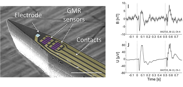
Figure 7 Left: Scanning Electron Microscopy of a magnetrode probe, comprising GMR sensors on a needle-shaped silicon substrate. Scalebar: 100 μm. Right: (Top) Magnetic recordings on the GMR sensor of the Evoked Response to a visual stimulus as a function of time. (Bottom) Electrical recordings from a tungsten electrode on the same stimulus. Exposure to the stimulus is indicated by the vertical dashed lines. Signals are averaged 2000 times.
Perspectives of these local magnetic recordings are to target the measurement of single events and of single cell responses, in particular through neuronal action potential detection which would lead to a spatially and time-resolved mapping of the neural currents running inside neurons.
F. Barbieri et al. Scientific Reports 6, (2016)
L. Caruso et al. Neuron , Volume 95 , Issue 6 , 1283 – 1291(2017)
Neuronal activity generates ionic flows and thereby both magnetic fields and electric potential differences,
i.e., voltages. Voltage measurements are widely used but suffer from isolating and smearing properties of tissue between source and sensor. They are blind to ionic flow direction, and reflect the difference between two electrodes, complicating interpretation. Magnetic field measurements could overcome these limitations but have been essentially limited to magnetoencephalography (MEG), using centimeter-sized, helium-cooled extracranial sensors. Here, we report on in vivo magnetic recordings of neuronal activity from visual cortex of cats with magnetrodes, specially developed needle-shaped probes carrying micron-sized, non-cooled magnetic sensors based on spin electronics. Event-related magnetic fields inside the neuropil were on the order of several nanoteslas, informing MEG source models and efforts for magnetic field measurements through MRI. Though the signal-to-noise ratio is still inferior to electrophysiology, this proof of concept demonstrates the potential to exploit.
Currents circulating in excitable cells like neurons or nerve fibers may be measured by the radiated magnetic field. At the organ level, these magnetic fields can be detected by non-invasive experiments using highly sensitive magnetometers such as SQUIDS, atomic magnetometers or mixed sensors, the latter using spin electronics. This technique, called Magneto-Encephalography, allows measuring neuronal activity at a millisecond resolution and for collective response of population of typically 10000 neurons and more. To understand the genesis of the signals obtained in brain areas, it is relevant to investigate the fields generated at the level of one or few cells. This requires small and sensitive field sensors, operating at physiological temperatures, which has long been out of reach from existing technologies.
Spin electronics allow now developing small sized and very sensitive magnetometers, reaching the sub-nanotesla field range on micron-size sensors. These devices operate from low temperature to hundreds of °C, so they can be used at physiological temperature. Furthermore, spin electronics sensors, based on thin film technology, can be deposited on silicon or glass substrates which can be shaped in needle-type devices to allow penetration in tissues with reduced damages.
We have designed and fabricated magnetic sensors called magnetrodes, as a magnetic equivalent of electrodes, to probe locally the information transmission of excitable cells. These sharp probes contain GMR elements in embodiment compatible with recordings within tissues.
After measuring magnetic action potential in vitro in a muscle with a flat probe , we have realised the first in vivo experimental measure of the magnetic signature of local field potentials in the cat’s visual cortex . The experiments have been conducted in collaboration with the Ernst Strüngmann Institute in Frankfurt. It has paved a new way to a local description of electrical activity, without direct contact to the cell and which allows accessing not only the amplitude of the activity but also its direction of propagation, at any depth within the tissues.
Figure 7 Left: Scanning Electron Microscopy of a magnetrode probe, comprising GMR sensors on a needle-shaped silicon substrate. Scalebar: 100 μm. Right: (Top) Magnetic recordings on the GMR sensor of the Evoked Response to a visual stimulus as a function of time. (Bottom) Electrical recordings from a tungsten electrode on the same stimulus. Exposure to the stimulus is indicated by the vertical dashed lines. Signals are averaged 2000 times.
Perspectives of these local magnetic recordings are to target the measurement of single events and of single cell responses, in particular through neuronal action potential detection which would lead to a spatially and time-resolved mapping of the neural currents running inside neurons.
F. Barbieri et al. Scientific Reports 6, (2016)
L. Caruso et al. Neuron , Volume 95 , Issue 6 , 1283 – 1291(2017)